Neuroblastoma

What is a Neuroblastoma?

Neuroblastoma is a cancer that starts in certain very early forms of nerve cells, most often found in an embryo or fetus. (The term neuro refers to nerves, while blastoma  refers to a cancer that starts in immature or developing cells). This type of cancer occurs most often in infants and young children.

The types of cancers that develop in children are often different from the types that develop in adults. To understand neuroblastoma, it helps to know about the sympathetic nervous system, which is where these tumors start.

The sympathetic nervous system

The brain, spinal cord, and the nerves that reach out from them to all areas of the body are all part of the nervous system. The nervous system is needed for thinking, sensation, and movement, among other things.

The main cells that make up the nervous system are called nerve cells or neurons. These cells interact with other types of cells in the body by releasing tiny amounts of chemicals (hormones). This is important, because neuroblastoma cells often release certain chemicals that can cause symptoms

Neuroblastomas

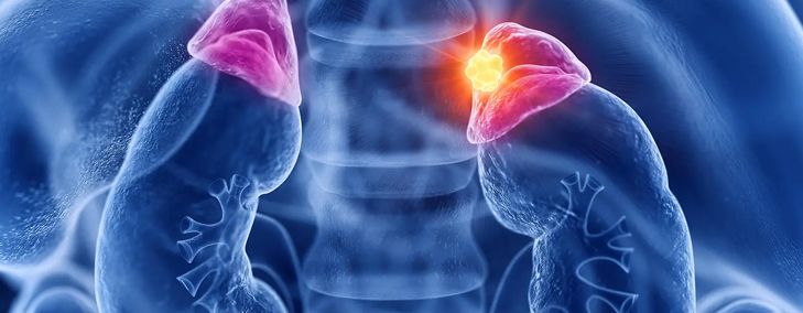
Neuroblastomas are cancers that start in early nerve cells (called neuroblasts) of the sympathetic nervous system, so they can be found anywhere along this system.

    • Most neuroblastomas begin in the abdomen, either in an adrenal gland or in sympathetic nerve ganglia.
    • Most of the rest start in sympathetic ganglia near the spine in the chest or neck, or in the pelvis.
    • Rarely, a neuroblastoma has spread so widely by the time it is found that doctors can’t tell exactly where it started.

Some neuroblastomas grow and spread quickly, while others grow slowly. Sometimes, in very young children, the cancer cells die for no reason and the tumor goes away on its own. In other cases, the cells sometimes mature on their own into normal ganglion cells and stop dividing (which makes the tumor a benign ganglioneuroma – see below).

Not all childhood autonomic nervous system tumors are malignant (cancerous). Some tumors are benign (non-cancerous), and some can have both benign and cancer cells within the same tumor.

  • Ganglioneuroma is a benign (non-cancerous) tumor made up of mature ganglion and nerve sheath cells.
  • Ganglioneuroblastoma is a tumor that has both malignant and benign parts. It contains neuroblasts (immature nerve cells) that can grow and spread abnormally, similar to neuroblastoma, as well as areas of more mature tissue that are similar to ganglioneuroma.

If a child is thought to have one of these tumors, it is usually removed by surgery and looked at carefully with a microscope to see if it has areas of cancer cells (which would make it a ganglioneuroblastoma). If the final diagnosis is ganglioneuroma, no other treatment is needed. If it’s found to be a ganglioneuroblastoma, it’s treated the same way as a neuroblastoma.

Tests for Neuroendocrine Tumors:

Neuroblastoma can start in different places in the body. Neuroblastoma cells can also sometimes release chemicals called hormones, which can affect other parts of the body. Because of this, neuroblastoma can cause many different signs and symptoms. Some of the more common symptoms can include:

  • Lump or swelling in the child’s belly that doesn’t seem to hurt
  • Swelling in the legs or in the upper chest, neck, and face
  • Problems with breathing or swallowing
  • Weight loss
  • Not eating or complaining about feeling full
  • Problems with bowel movements or urinating
  • Pain in bones
  • Lumps or bumps under the skin, which may appear blue
  • Drooping eyelid and small pupil (the black area in the center of the eye) in one eye
  • Problems being able to feel or move parts of the body
  • Eyes that appear to bulge and/or bruising around the eyes

Signs and symptoms might be different depending on where the tumor is, how large it is, how far it has spread, and if the tumor makes hormones.

Tests for Neuroblastoma:

If a neuroblastoma is found, other tests will then be needed to learn more about it.

Medical history and physical exam

If the history and exam suggest a child might have a neuroblastoma (or another type of tumor), more tests will be done. These could include:

  • Blood and urine tests
  • Imaging tests
  • Biopsies
Blood and urine catecholamine tests
  • Homovanillic acid (HVA)
  • Vanillylmandelic acid (VMA)

Neuroblastoma cells also often make these catecholamines, so these same metabolites can be get an idea of how well it is working.

Other blood and urine tests: If neuroblastoma is suspected or has been found, your child’s doctor will probably order blood tests to check blood cell counts, liver and kidney function, and the balance of salts (electrolytes) in the body. A urinalysis (urine test) may also be done to help check kidney function.

Imaging tests

Most children who have or might have neuroblastoma will get one or more of these tests, but they might not need all of them.

Radiation therapy:

Radiation therapy may be used in patients in whom tumour is localized but may not be easily operable.

Ultrasound (sonogram)
Magnetic resonance imaging (MRI)
Computed tomography (CT or CAT) scan
CT-guided needle biopsy
MIBG scan

This test is often an important part of finding out how far a child’s neuroblastoma has spread.

For this test, a form of the chemical meta-iodobenzylguanidine (MIBG) that contains a small amount of radioactive iodine is injected into the blood. MIBG is similar to norepinephrine, a hormone made by sympathetic nerve cells, and in most patients it will attach to neuroblastoma cells anywhere in the body. Between 1 and 3 days later, the body is scanned with a special camera to look for areas that picked up the radioactivity. This helps doctors know where the neuroblastoma is and if it has spread to the bones and/or other parts of the body.

MIBG scans can be repeated after treatment to see if the tumors are responding well. It is also good to know if the tumor takes up the MIBG because in some cases, this radioactive molecule can be used at higher doses to treat the neuroblastoma.

  • Bone scan
  • Positron emission tomography (PET) scan
  • X-rays
  • Biopsies

During a biopsy, a doctor removes one or more pieces (samples) from the tumor for testing.
There are 2 main types of biopsies:

  1. Incisional (open or surgical) biopsy
  2. Needle (closed) biopsy

Once the biopsy samples have been removed, they are sent to a lab, where they are viewed under a microscope by a pathologist (a doctor with special training in identifying cancer cells). Special lab tests are often done on the samples as well to show if the tumor is a neuroblastoma.

Treatment options for Neuroblastoma:
  • Neuroblastoma Surgery
  • Chemotherapy
  • Radiation Therapy
  • Targeted therapy
  • MIBG therapy
  • High-dose Chemotherapy and Stem Cell Transplant
  • Retinoid Therapy
  • Immunotherapy

    Make Appointment

    Get in touch with us today +91 8130168644